2015
Abstract
Since the enactment of Title IX (1972), rates of female participation in sports have grown exponentially. However, despite these growing numbers, women are infrequently asked about risk factors for and consequences of concussion in the ambulatory care setting or during well-woman visits. As well, concussion in women and men usually are not differentiated when considering presentation and adverse outcomes. Data are emerging that suggest concussion not only has long-term health implications but also that the effect of this injury in women compared with men is related to physical differences, cognitive differences (particularly in the verbal, quantitative, and visuospatial domains) and differences in the hormonal milieu. This paper serves as a review of the literature on concussion in the female athlete on PubMed was conducted, with attention to both clinical studies and diagnostic studies. Results suggest that sex and gender contribute to concussion prevalence and severity, presenting symptoms, and recovery. For example, women may present with more drowsiness and sensitivity to noise after concussion than men. As well, increased surveillance appears to be warranted despite obvious behavioral recovery. Results of a longitudinal functional magnetic resonance imaging study in athletes with concussion suggest that neural recovery lags behind behaviorally assessed recovery, and alterations in white matter fiber tracts over the 2 months after injury have been demonstrated. Screening templates are available, and consideration of incorporation of relevant questionnaires into the electronic health record as well as the potential role of imaging will be discussed. In conclusion, inquiry about concussion should be considered a part of the well-woman visit for all patients, especially those engaged in at-risk contact sports. The gynecologic visit and examination should include questions about head injury from sports involvement and a prior diagnosis of concussion.
2014
Abstract
Avoiding recurrent injury in sports-related concussion (SRC) requires understanding the neural mechanisms involved during the time of recovery after injury. The decision for return-to-play is one of the most difficult responsibilities facing the physician, and so far this decision has been based primarily on neurological examination, symptom checklists, and neuropsychological (NP) testing. Functional magnetic resonance imaging (fMRI) may be an additional, more objective tool to assess the severity and recovery of function after concussion. The purpose of this study was to define neural correlates of SRC during the 2 months after injury in varsity contact sport athletes who suffered a SRC. All athletes were scanned as they performed an n-back task, for n = 1, 2, 3. Subjects were scanned within 72 hours (session one), at 2 weeks (session two), and 2 months (session three) post-injury. Compared with age and sex matched normal controls, concussed subjects demonstrated persistent, significantly increased activation for the 2 minus 1 n-back contrast in bilateral dorso- lateral prefrontal cortex (DLPFC) in all three sessions and in the inferior parietal lobe in session one and two (a £ 0.01 corrected). Measures of task performance revealed no significant differences between concussed versus control groups at any of the three time points with respect to any of the three n-back tasks. These findings suggest that functional brain activation differences persist at 2 months after injury in concussed athletes, despite the fact that their performance on a standard working memory task is comparable to normal controls and normalization of clinical and NP test results. These results might indicate a delay between neural and behaviorally assessed recovery after SRC.
Abstract
2013
Abstract
2011
Abstract
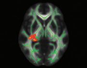
Recognizing and managing the effects of cerebral concussion is very challenging, given the discrete symp- tomatology. Most individuals with sports-related concussion will not score below 15 on the Glasgow Coma Scale, but will present with rapid onset of short-lived neurological impairment, demonstrating no structural changes on traditional magnetic resonance imaging (MRI) and computed tomography (CT) scans. The return-to- play decision is one of the most difficult responsibilities facing the physician, and so far this decision has been primarily based on neurological examination, symptom checklists, and neuropsychological (NP) testing. Dif- fusion tensor imaging (DTI) may be a more objective tool to assess the severity and recovery of function after concussion. We assessed white matter (WM) fiber tract integrity in varsity level college athletes with sports- related concussion without loss of consciousness, who experienced protracted symptoms for at least 1 month after injury. Evaluation of fractional anisotropy (FA) and mean diffusivity (MD) of the WM skeleton using tract- based spatial statistics (TBSS) revealed a large cluster of significantly increased MD for concussed subjects in several WM fiber tracts in the left hemisphere, including parts of the inferior/superior longitudinal and fronto- occipital fasciculi, the retrolenticular part of the internal capsule, and posterior thalamic and acoustic radiations. Qualitative comparison of average FA and MD suggests that with increasing level of injury severity (ranging from sports-related concussion to severe traumatic brain injury), MD might be more sensitive at detecting mild injury, whereas FA captures more severe injuries. In conclusion, the TBSS analysis used to evaluate diffuse axonal injury of the WM skeleton seems sensitive enough to detect structural changes in sports-related con- cussion.

